What is an angiogram?
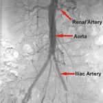
An angiogram or arteriogram is an X-ray test that uses dye to demonstrate the arteries. Arteries are invisible to X-ray so the only way they can be seen is by filling them with dye. The correct name for the dye is ‘contrast’, and it is the iodine it contains that is visible on X-ray.
Angiograms are performed or supervised by a consultant radiologist in the X-ray department. Most vascular surgeons do not perform the angiogram themselves.
There are three ways in which an angiogram may be performed:
1. Digital subtraction angiography (DSA)
2. Computed tomography angiography (CTA)
3. Magnetic resonance angiography (MRA)
1. Digital subtraction angiography (DSA)
DSA is now regarded as the “conventional” way of getting pictures of the arteries. The site of the area to be investigated is numbed with a local anaesthetic (this usually stings as the anaesthetic is injected). The artery to be investigated is then injected with contrast, which then outlines the arteries beyond.
As arteries are under high pressure it is important not to allow this to drop. Once the contrast has been injected and the needle removed, pressure is applied to the puncture site for 10 – 20 minutes or a sealing device is inserted to seal the hole.
The X-ray machine used to acquire the pictures is usually a large “C” shaped arm which curves around you. It is not claustrophobic, and you will be able to watch the procedure on the monitor screens above you.
The procedure usually takes about 20 minutes. DSA is sometimes performed as a day case, and sometimes involves a one-night stay in hospital.
2. Computed Tomography Angiogram (CTA)
As CT scanners have become more sophisticated and quicker, they can now be used to generate an angiogram. This can be displayed as a 3-dimensional image on a computer screen as shown on the right. This picture shows the circulation from the diaphragm down to the ankles.
Like a DSA, contrast is injected, but instead of injecting into the artery, the injection is made into a vein in the arm. The arteries anywhere in the body can then be outlined. Preparation for the procedure involves putting a drip in the arm.
A CT scan involves lying on a narrow table which moves through the scanner as the images are acquired. The images only take a few seconds to generate. The procedure is very quick, and can always be performed as a day case. Like angiography it is not claustrophobic.
3. Magnetic Resonance Angiogram (MRA)
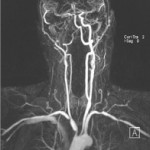
An MR scan uses a magnetic field to generate images. Like CT it can create 3-dimensional images on a computer. Contrast (gadolinium) is injected into a vein in your arm. Any arteries in the body can then be viewed as a 2 or 3-dimensional image. The image on a computer can be rotated and viewed from any angle.
An MR scan involves lying on a table inside a narrow tube. This can cause claustrophobia for some people.
The procedure is very quick and can always be performed as a day case.
How will the angiogram be done?
Your consultant will discuss this and advise on the best method for you. Before the angiogram is done, you will have a blood test to check your blood count, blood clotting and kidney function, and you will be asked to sign a consent form.
On the day you should take all your medication as usual. If you are on Warfarin or Clopidogrel (Plavix), they normally need to be stopped. Make sure your consultant knows if you are on either of these drugs.
Angiograms are nearly always done under local anaesthetic while you are awake. Very occasionally and under exceptional circumstances, a general anaesthetic is required.
What are the risks?
Angiography is very safe. More than 95 in every 100 will have no complications at all.
Allergy to the contrast is a problem for some patients, and can be overcome either by giving steroids at the time of injection or by using MRA, which doesn’t involve the injection of iodine.
Claustrophobia during MRA can be a problem for some patients, and if this is the case DSA or CTA can usually be chosen instead.
In about 5 in 100 DSA cases there may be bleeding from the arterial puncture site. If this happens, it will be within a few hours, and can usually be stopped. In extreme cases a small operation to repair the artery is required. Following arterial puncture for DSA a small number of patients develop a false aneurysm. A false aneurysm is a cavity beneath the skin connecting to the artery by its puncture hole. There is usually pain over the aneurysm. A false aneurysm can be treated by compression, injection of thrombin (which causes it to clot and disappear) or by surgery to repair the hole in the artery.
Injection of contrast can occasionally upset kidney function, particularly if there is already a degree of kidney damage. A routine pre-procedure blood test will always be done, and will show if this is the case. Special measures can be taken to avoid this complication.
What happens afterwards?
Everyone gets a bit of bruising where the artery (or vein) was punctured. It is variable and unpredictable. The bruising will normally be sore and uncomfortable for 7-10 days, and the visible bruising may take up to 4 weeks to resolve. Persistent pain at an arterial puncture site may indicate a false aneurysm. You should contact your GP or consultant.
After 24 hours, unless you are told otherwise, there are no restrictions on your level of activity. Driving, work, lifting, bathing and sport are all OK.
Information and advice about vascular health.
Whilst we make every effort to ensure that the information contained on this site is accurate, it is not a substitute for medical advice or treatment, and the Circulation Foundation recommends consultation with your doctor or health care professional.
The Circulation Foundation cannot accept liability for any loss or damage resulting from any inaccuracy in this information or third party information such as information on websites to which we link.
The information provided is intended to support patients, not provide personal medical advice.


